Kidney Regeneration & Frontiers: Botanicals, HBOT, 3D Printing & Xenotransplantation
Patients with end-stage renal disease (ESRD) frequently ask whether lost kidney function can ever be restored. The internet is filled with claims about herbal cures, hyperbaric chambers, and stem cell injections. Here is the unvarnished science of what is biologically possible, what carries lethal risks, and where genuine biomedical breakthroughs are happening.
Why adult kidneys do not naturally regenerate in ESRD
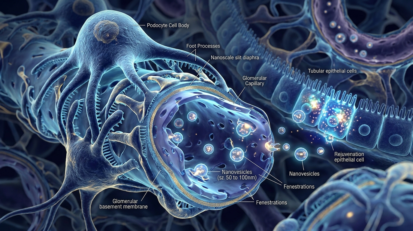
Unlike the liver, which can regrow after substantial tissue loss, the adult human kidney has strictly limited regenerative capacity once end-stage damage is established.
The kidney's primary filtration barrier relies on podocytes—specialized epithelial cells with intricate interdigitating foot processes and microscopic slit diaphragms (4 to 11 nanometers wide). Podocytes are terminally differentiated and post-mitotic; once they detach or undergo apoptosis, they cannot divide to replace missing cells.
While surviving tubular cells can regenerate after acute tubular necrosis (ATN), chronic kidney disease leads to irreversible microvascular rarefaction (loss of peritubular capillaries) and progressive tubulointerstitial fibrosis. Driven by chronic TGF-beta1 signaling, the renal tissue is replaced by dense, cross-linked Type I and Type III collagen that chokes off residual blood flow. True regeneration requires both dissolving this fibrous scar and reconstructing over 800,000 microscopic filtration units connected to high-pressure arterial circulation.
Herbal medicine & botanicals: biological evidence vs nephrotoxicity risks
Several botanical compounds have demonstrated measurable anti-inflammatory and anti-fibrotic activity in preclinical and early clinical CKD research, but they carry severe hazards for patients with ESRD.
- Astragalus membranaceus (Huang Qi): Active fractions (astragaloside IV and cycloastragenol) downregulate TGF-beta1/Smad signaling, induce the Nrf2 antioxidant cascade, and attenuate podocyte transdifferentiation. Cochrane systematic reviews show modest reductions in proteinuria in CKD Stages 2–4, but it cannot rebuild dead glomeruli.
- Cordyceps sinensis (CS-4): Stimulates adenosine A1/A2A receptors, attenuates TLR4/NF-kappaB pro-inflammatory signaling, and helps mitigate calcineurin inhibitor nephrotoxicity in transplant models.
- Rheum officinale (Da Huang / Emodin): Acts through the gut-kidney axis by binding uremic toxins (such as indoxyl sulfate and p-cresyl sulfate) in the colon and suppressing tubular fibronectin deposition.
- Salvia miltiorrhiza (Dan Shen / Tanshinone IIA): Promotes endothelial nitric oxide synthase (eNOS) phosphorylation, improving microcirculatory perfusion and blunting ischemia-reperfusion injury.
- CRITICAL DANGER — The 'Fang Ji' Confusion & Belgian Tragedy: A notorious case of herbal nephrotoxicity occurred in the early 1990s when slimming pills in Belgium mistakenly substituted non-nephrotoxic Han Fang Ji (Stephania tetrandra, Menispermaceae) with Guang Fang Ji (Aristolochia fangchi, Aristolochiaceae). Because the dried roots appear virtually indistinguishable and shared the common name 'Fang Ji', over 100 young patients developed rapid-onset extensive renal fibrosis, end-stage renal failure, and high rates of upper tract urothelial cancer (UTUC).
- The 'Mu Tong' & Balkan Contaminations: The exact same deadly substitution plagued Mu Tong, where toxic Guan Mu Tong (Aristolochia manshuriensis) was substituted for safe Chuan Mu Tong (Clematis armandii) or Akebia quinata. Similarly, Balkan Endemic Nephropathy (BEN) was traced to Aristolochia clematitis seeds contaminating local wheat crops and bread flour.
- Molecular Mechanism of Aristolochic Acid Nephropathy (AAN): Aristolochic acid I (AAI) forms covalent DNA adducts (7-(deoxyadenosin-N6-yl)aristolactam I, dA-AAI) that cause a signature, irreversible A:T to T:A transversional mutation in the TP53 tumor suppressor gene, simultaneously inducing acute tubular apoptosis, microvascular devastation, and rapid acellular fibrotic destruction of the kidney parenchyma.
- Why AAN is Deceptive: Aristolochic acid nephropathy presents with an eerie absence of cellular inflammation on kidney biopsy (acellular interstitial fibrosis) and minimal proteinuria, meaning patients can progress silently to irreversible ESRD without the typical warning signs of nephritis.
- Electrolyte and Heavy Metal Hazards: In anuric or oliguric dialysis patients, unstandardized herbal decoctions frequently contain fatal concentrations of potassium and toxic heavy metals. Any botanical product must be third-party certified pure and approved by your renal team.
Hyperbaric Oxygen Therapy (HBOT): Gas physics & tissue hypoxia
Chronic tubulointerstitial hypoxia is considered the final common pathway driving progressive renal fibrosis. Sclerotic glomeruli restrict blood flow into post-glomerular capillaries, depriving tubules of oxygen.
- Physiological mechanism: Breathing 100% oxygen at 2.0 to 2.4 atmospheres absolute (ATA) dissolves up to 6.0 mL of oxygen per deciliter of plasma (a twenty-fold increase over room air), independent of hemoglobin delivery.
- The hyperoxic-hypoxic paradox: Periodic hyperbaric pulses trigger bone marrow mobilization of CD34+ endothelial progenitor cells (EPCs) and transiently stimulate VEGF-mediated capillary sprouting without sustained ischemia.
- Approved clinical indications: HBOT is clinically approved and effective for treating severe non-healing ischemic wounds and calciphylaxis in ESRD patients.
- Clinical limitation in atrophic kidneys: In end-stage kidneys that have shrunken below 8 cm with extensive calcification, the cellular architectural templates are gone; hyperbaric oxygen cannot resurrect deleted nephrons.
- Oxidative stress caution: ESRD patients suffer from chronic glutathione depletion. Excessive hyperoxia generates reactive oxygen species (ROS) and lipid peroxidation unless properly monitored and counterbalanced with physician-guided antioxidant defenses.
Stem cell therapies & exosomes: why MSCs don't become kidneys
One of the most persistent myths in regenerative medicine is that injecting Mesenchymal Stem Cells (MSCs) will cause them to home into the kidney and differentiate into new nephrons.
- The paracrine secretome: Extensive cell-fate tracking has proven that over 95% of stem cell therapeutic benefits come from the molecules they secrete, not cell replacement.
- Extracellular vesicles (Exosomes): Acellular 30 to 150 nm vesicles derived from umbilical cord or bone marrow MSCs carry microRNAs (such as miR-let-7c and miR-29b) that downregulate collagen transcription and activate matrix metalloproteinase-9 (MMP-9) to digest soft interstitial scars.
- Klotho hormone therapy: Produced primarily in healthy renal tubules, Klotho is the body's master anti-fibrotic hormone. In ESRD, circulating Klotho is nearly absent. Experimental administration of recombinant Klotho or Klotho-upregulating peptides suppresses Wnt/beta-catenin signaling, halts vascular calcification, and protects cardiac muscle.
- Clinical warning: Commercial overseas clinics offering direct stem cell injections for kidney failure are unregulated, unapproved by the FDA, and pose severe risks of pulmonary embolism, bacterial infection, and tumor formation.
The engineering reality of 3D bioprinting & decellularized ECM
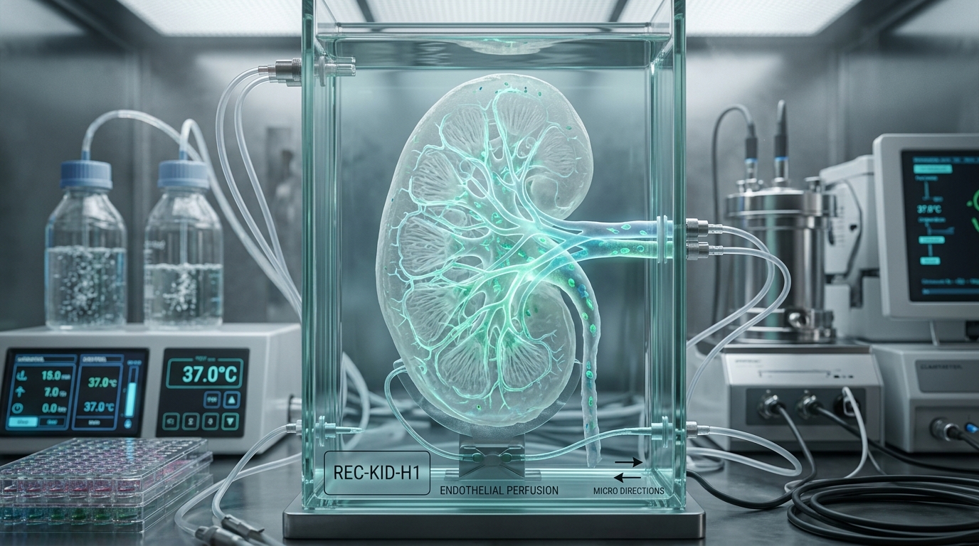
3D bioprinting of functional vascularized kidneys remains one of biomedical engineering's most daunting frontiers.
- The microstructural bottleneck: A human kidney contains approximately one million nephrons. Mechanical bioprinters typically deposit cells at 50 to 100 micrometer resolution, whereas glomerular capillary loops (5–10 micrometers) and podocyte slit diaphragms (4–11 nanometers) require molecular self-assembly that cannot be mechanically sprayed.
- Decellularized whole-organ scaffolds: Washing donor porcine kidneys with detergents produces a pristine collagen-elastin vascular tree. However, 100% complete re-endothelialization of every microscopic capillary is mandatory. If even a fraction of a percent of bare collagen is exposed to flowing human blood, immediate platelet aggregation and catastrophic vascular thrombosis occur upon reperfusion.
- Realistic timeline: While 3D bioprinted proximal tubule models (such as organ-on-a-chip platforms) are currently revolutionizing pharmaceutical toxicology, fully functional implantable 3D bioprinted kidneys remain 12 to 20 years away from clinical practice.
The two near-term clinical breakthroughs: Bioartificial kidneys & Xenotransplantation
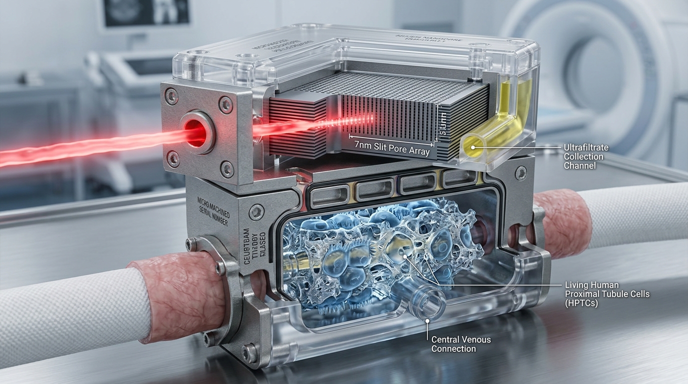
While cellular 3D bioprinting remains long-term, two technological avenues are advancing rapidly in human clinical medicine.
- The Bioartificial Implantable Kidney (The Kidney Project - UCSF & Vanderbilt): Co-directed by Dr. Shuvo Roy and Dr. William Fissell, this two-stage device pairs a semiconductor silicon nanopore membrane (SNM, pore size ~7 nanometers) with a bioreactor of living human renal tubule cells. It operates entirely on the patient's arterial blood pressure—requiring zero pumps, zero external dialysate, and no systemic immunosuppressive drugs because the silicon membrane immunoisolates the tubule cells.
- CRISPR-Edited Porcine Xenotransplantation: Scientists at eGenesis, Revivicor, and academic centers have engineered miniature swine with up to 69 genetic modifications—knocking out 3 carbohydrate xenoantigens (GGTA1, B4GALNT2, CMAH) to prevent hyperacute rejection, adding 7 human complement and antithrombotic transgenes, and inactivating 59 porcine endogenous retrovirus (PERV) loci.
- Historic human milestones: Living human transplantations performed at Massachusetts General Hospital and NYU Langone demonstrated immediate urine production, successful creatinine clearance, and restoration of eGFR above 50 mL/min.
- Novel immunosuppression: Costimulation blockade therapies (such as anti-CD40 monoclonal antibodies) protect xenografts from rejection without the nephrotoxic damage caused by traditional calcineurin inhibitors.
Actionable questions for your nephrologist
- What is my current residual kidney function (eGFR and 24-hour urine volume), and how can we protect it?
- Are there open clinical trials (such as high-volume HDF, novel tolerance protocols, or expanded-access xenotransplantation) for which I meet the eligibility criteria?
- Can we safely review every supplement, tea, and botanical I consume against my latest electrolyte panel to eliminate potassium and nephrotoxicity risks?
- If I have chronic ischemic wounds or poor circulation, would adjuvant hyperbaric oxygen therapy be safe and medically indicated for my condition?